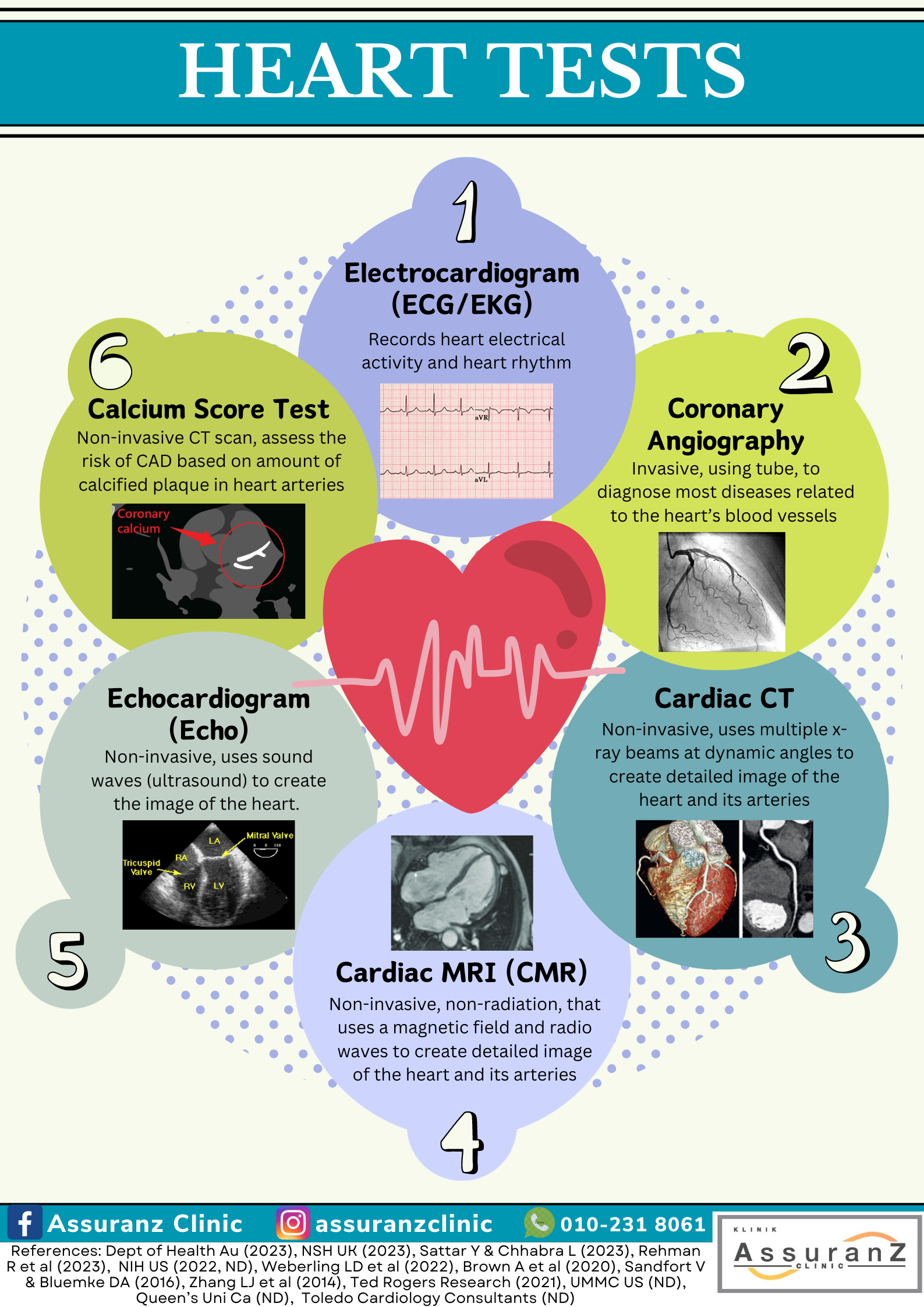
Electrocardiogram (ECG/EKG) / 心电图
An ECG test is a non-invasive test to monitor the electrical activity of the heart. It does not show the image of the heart. It is used as a screening tool and as an initial clinical diagnostic method for patients suspected to have heart-related problems. An ECG can help diagnose abnormal heart electrical activity and heart rhythm, heart attack, inflammation, inadequate blood supply to heart and cardiac arrest. It is also used to monitor treatments for heart-related conditions such as medication and cardiac implantable devices.
心电图(ECG)是一项快速且无痛的检查,用于监测心脏的电信号。它并不显示心脏的图像,而是作为筛查工具和初步临床诊断方法,帮助识别疑似心脏问题的患者。心电图可帮助诊断心脏电活动异常、心律失常、心脏病发作、心脏炎症、心脏供血不足及心脏骤停等问题。它还用于监测心脏相关疾病的治疗效果,包括药物治疗和心脏植入设备的使用。
- Resting ECG / 静息心电图
Resting ECG is used to record the heart’s electrical activity during rest (lying down with no movement).
静息心电图用于记录心脏处于休息状态下(躺着且没有活动时)的电信号。
- Stress Test / 运动负荷试验
Exercise stress test is to determine the heart’s electrical activity in response to external stress, that is physical activity. This involves having an ECG reading taken while cycling on stationary bike or walking on a treadmill.
运动负荷试验用于评估心脏在外部压力下(即体力活动)的电信号反应。这项测试通常是在患者骑静止自行车或走在跑步机时,进行心电图监测。
- Ambulatory ECG / 动态心电图
Ambulatory ECG, as the name suggests, is a small portable ECG which is used to record the electrical wave of the heart for a certain period, typically between 24 hours to 7 days, as decided by the medical practitioner. This ECG is to evaluate symptoms and abnormalities that only occur occasionally which cannot be detected by the normal ECG.
动态心电图,顾名思义,是一种小型便捷式心电图设备,在医生的指定的时间内(通常为24小时至7天),用于记录心脏的电信号。该检查旨在评估那些偶尔发生、常规心电图无法检测到的症状和异常情况。
Imaging / 影像检查
Imaging tests are tests that can provide detailed images of the area inside the body or of an organ. The images can be used to determine abnormal heart conditions such as artery occlusion, abnormality of the valve, enlarged heart size, congenital heart disease et cetera. They include the application of different energy forms, for instance, high energy radiation (x-ray), high energy sound waves (ultrasound), radio waves and radioactive matters.
影像检查是一项能够提供身体内部区域或器官详细图像的检查。这些图像可用来判断心脏异常情况,如动脉堵塞、心脏瓣膜异常、心脏扩大、先天性心脏病等。影像检查包括使用不同形式的能量,例如高能辐射(x射线)、高能声波(超声波)、无线电波及放射性物质。
- Coronary Angiography / 冠状动脉造影
Coronary angiography, also known as coronary catheterization, is the diagnostic gold standard for most diseases related to the heart’s blood vessels such as coronary artery disease (CAD). It is an invasive technique involving a tube (catheter) guided through a blood vessel to the heart. However, the current guidelines recommend non-invasive testing prior to invasive procedures.
冠状动脉造影,也称为冠状动脉导管插入术,是诊断大多数与心脏血管相关的疾病,如冠状动脉疾病(CAD)的金标准方法。这是一项侵入性的检查技术,通过将导管插入血管并引导心脏进行检查。然而,目前的指南建议在进行侵入性操作前,先进行非侵入性检查。
- Cardiac computed tomography (Cardiac CT) / 心脏断层扫描(心脏CT)
Cardiac CT is a non-invasive technique which uses multiple x-ray beams at dynamic angles to create image of the heart. The cardiac CT angiogram (CCTA) is a type of specialized CT scan that can be used to diagnose of CAD. Combining CT scan with contrast ‘dye’, it can picturize the image of the heart, including of the arteries. It is the low-risk alternative to coronary angiogram for stable patients at risk of CAD. There is exposure to radiation for this procedure, but it is minimal.
心脏CT是一种通过多角度的X射线束生成心脏图像的非侵入性影像检查技术。心脏CT血管造影(CCTA)是一种专门用于诊断冠状动脉疾病(CAD)的CT扫描。通过将CT扫描与造影剂的结合使用,它能够清晰地呈现心脏及其动脉的图像。对于那些处于冠状动脉疾病风险中的稳定患者,心脏CT是冠状动脉造影的低风险替代方案。虽然该过程会有辐射,但辐射量非常低。
- Cardiac Magnetic Resonance Imaging (CMR) / 心脏磁共振成像(CMR)
Cardiac MRI is a non-invasive, non-radiation imaging technique that uses a magnetic field and radio waves to create detailed image of the heart and its arteries. However, currently, CCTA is a more superior technique.
心脏磁共振成像是一种非侵入性、无辐射成像技术。它利用磁场和无线电波来呈现心脏和其动脉的详细图像。然而,目前心脏CT血管造影(CCTA)是一种更先进的技术。
- Echocardiogram (Echo)/Cardiac Ultrasound
超声心电图(Echo)/ 心脏超声波
Echocardiogram is a non-invasive test that uses sound waves (ultrasound) to create the image of the heart. A Doppler echo, which is a type of echo can also show how well the blood flows within the chambers and valves. While this test is convenient, rapid and efficient, in certain cases, other organs and body tissues such as the lungs, ribs, and air may affect the sound waves and echoes from producing clear image of the heart.
超声心电图是一种非侵入性检查。它使用声波(超声波)来创建心脏的图像。多普勒超声是一种超声检查类型,它还可以显示血液在心脏腔室和瓣膜内流动的情况。虽然这种检查既方便又快速高效,但在某些情况下,肺部、肋骨和空气等其他器官和身体组织可能会影响声波和回声,从而使心脏图像不够清晰。
- Coronary Calcium Scan/Calcium Score Test/Agatston Score
冠状动脉钙化扫描 / 钙化评分测试 / 阿加斯顿评分
Calcium score test is a non-invasive CT procedure used to assess the risk of CAD by calculating and categorizing the risk based on the amount of calcified plaque in the coronary arteries observed. This scoring does not directly diagnose any medical conditions, but it indicates that people with higher score are more likely to have cardiac event in comparison to other men or women of similar age.
钙化评分测试是一种非侵入性CT检查。它根据观察到的冠状脉钙化斑块的数量来计算和分类来评估冠状动脉疾病(CAD)的风险。此评分并不能直接诊断任何疾病,但它能表明与其他年龄相仿的男性或女性相比,分数较高的人更有可能患上心脏病。
Invasive: involving the introduction of instruments or other objects into the body or body cavities.
侵入性:将器械或其他物体引入身体或体腔。


